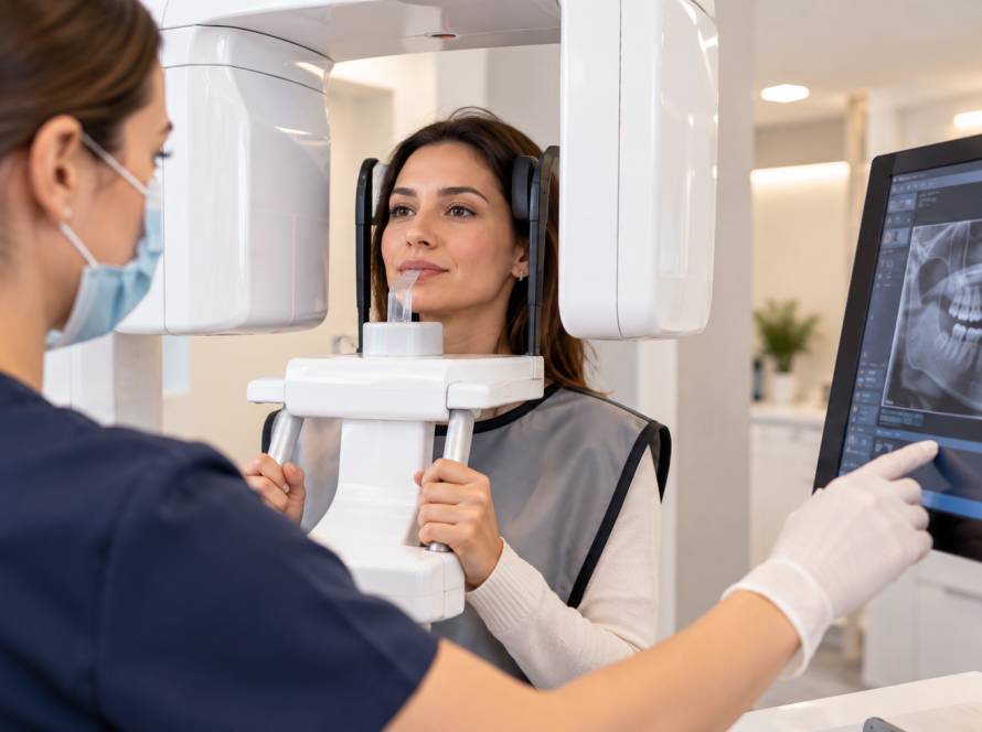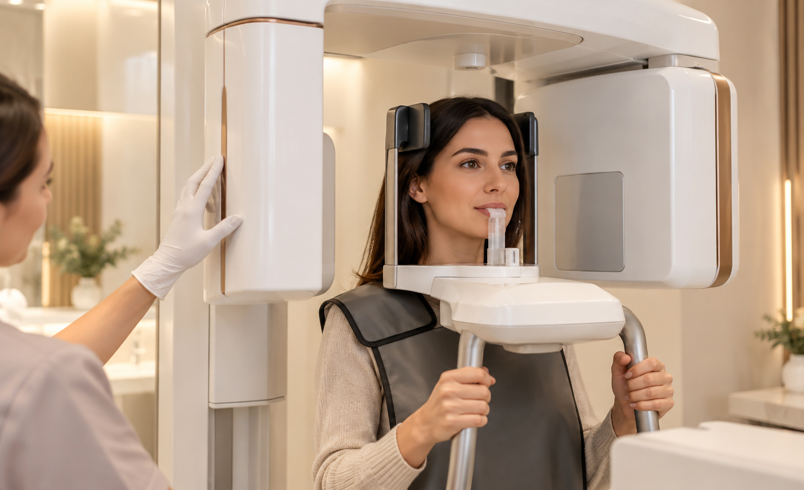
A panoramic dental X-ray is a two-dimensional image that shows the upper and lower jaws, teeth and several surrounding structures in one broad view. Unlike bitewing or periapical radiographs, the detector is outside the mouth and the imaging unit rotates around the head.
Its main advantage is coverage. A single image can provide a useful overview of tooth position, developing or impacted teeth, the jaws and some larger abnormalities. Its main limitation is that it is still a 2D image: structures can overlap, magnification and distortion can occur, and it does not provide the same fine local detail or three-dimensional anatomical information as other imaging methods.
A panoramic radiograph can therefore be very useful without being the right image for every dental question.
What a Panoramic Image Can Show
Panoramic imaging is helpful when a dentist needs to assess the mouth and jaws as a whole. Depending on the clinical question and image quality, it can show erupted and unerupted teeth, impacted or partially impacted wisdom teeth, general tooth development and eruption, broad jaw anatomy and some larger cystic or bony changes.
It can also help show general relationships between teeth and surrounding structures and identify areas that need more focused imaging.
The image is always interpreted alongside the clinical examination. A panoramic radiograph on its own is not a diagnosis.
The Limits of a Broad 2D View
Wide coverage comes with trade-offs. Panoramic radiography does not provide the highest-resolution view of every tooth, and small lesions can be obscured by overlapping structures or image geometry.
Bitewing radiographs are often better suited to detecting small caries lesions between posterior teeth, while periapical radiographs can provide more detailed local information around a particular tooth, its root and the surrounding apical area.
A panoramic image also cannot show true three-dimensional bone width or spatial relationships in the way CBCT can. A panoramic radiograph that looks normal therefore does not prove that every tooth and surrounding structure is free of disease.

Wisdom Teeth and Panoramic Imaging
When imaging is clinically indicated, a panoramic radiograph can provide a useful overview of wisdom-tooth eruption, angulation and the general relationship to the neighbouring second molar and surrounding jaw structures.
For many cases, that overview is sufficient for the initial assessment. If the image raises a specific concern about a close anatomical relationship and three-dimensional information could change management, CBCT may then be considered.
CBCT is not automatically required for every wisdom tooth.
Panoramic X-Rays in Implant Assessment
A panoramic radiograph can provide preliminary information about an edentulous area and the general jaw anatomy. It can help the dentist understand the broader setting before more detailed planning is undertaken.
Implant planning may, however, require assessment of bone width and the three-dimensional relationship to structures such as the mandibular canal or maxillary sinus. Those questions cannot be measured reliably from a 2D panoramic image alone.
The imaging approach depends on the stage of planning and the information that is actually needed.
Panoramic, Bitewing and Periapical Radiographs
These images are not simply larger and smaller versions of the same test.
Bitewings show the crowns of upper and lower teeth in a limited region and are particularly useful for selected caries and bone-level questions. Periapical radiographs show one tooth or a small group of teeth from crown to root tip together with nearby bone, making them useful for many endodontic and local periapical assessments.
Panoramic imaging covers a much wider anatomical area, but with less fine detail for each individual tooth. The best image is the one that answers the clinical question with appropriate diagnostic information.
Panoramic X-Ray and CBCT
Panoramic radiography is two-dimensional, while CBCT produces a three-dimensional volumetric dataset that can be viewed in multiple planes.
CBCT can provide more detailed spatial information, but greater detail does not mean it should be used routinely. Current radiographic principles emphasise justification: 3D imaging should be selected when the additional information is expected to affect diagnosis or treatment planning and lower-dimensional imaging cannot answer the question adequately.
How a Panoramic X-Ray Is Taken
The patient is positioned in the imaging unit so that the head and dental arches are aligned correctly. The X-ray source and detector then rotate around the head while the patient remains still.
Because the technique depends on positioning within a defined focal zone, movement or incorrect head position can reduce image quality. Exposure settings vary according to the equipment and patient factors, so a single universal exposure time or radiation dose should not be assumed for every machine.
Imaging Frequency Should Be Based on Clinical Need
Panoramic X-rays are not required at every dental visit. Age, symptoms, disease risk, previous images, oral-health history and the current treatment question all influence whether imaging is justified.
A previous radiograph can sometimes remain clinically useful. Repeating a panoramic image solely because a patient has attended a new clinic is not automatically necessary.
Dental Imaging at Güler Dent
Dental Radiology is one of the main clinical service areas at Güler Dent in Samsun, Turkey. The clinic has in-clinic tomography technology for situations in which three-dimensional imaging is clinically indicated.
The presence of tomography does not mean CBCT is used for every patient. The imaging method should match the diagnostic question and the findings from the clinical examination.