Dental X-Rays and CBCT in Turkey
Dental imaging supports diagnosis and treatment planning when information is needed beyond what can be seen during a clinical examination. It can help assess tooth roots, supporting bone, impacted teeth, lesions and anatomical relationships that may affect implant, endodontic, periodontal or surgical decisions.
Contents
ToggleThe correct image is not automatically the most detailed image. Panoramic and periapical radiographs are two-dimensional, while cone-beam computed tomography (CBCT) can provide three-dimensional information. Current radiographic guidance emphasises patient selection: imaging should be based on clinical need, and CBCT should not be used routinely when lower-dimensional imaging provides the information required.
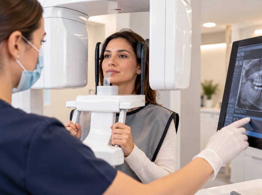
Güler Dent in Atakum, Samsun, Turkey has in-clinic tomography technology. CBCT is used according to clinical need rather than as a routine examination for every patient.
Why Are Dental X-Rays Used?
Radiographs can provide information about areas that cannot be directly examined visually. Depending on the clinical question, they may help assess decay, roots, bone levels, impacted teeth, root-area changes and anatomical structures relevant to treatment planning.
Imaging does not replace the clinical examination. Radiographic findings need to be interpreted together with symptoms, medical and dental history and clinical findings.
Panoramic, Periapical and CBCT Imaging
Panoramic Radiography
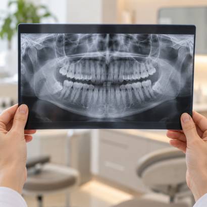
A panoramic radiograph is a two-dimensional image covering a broad area of the upper and lower jaws. It can be useful for general assessment, tooth eruption, impacted teeth and some surgical or orthodontic questions.
Because it is two-dimensional, structures can overlap and fine local detail may not always be sufficient.
Periapical Radiography
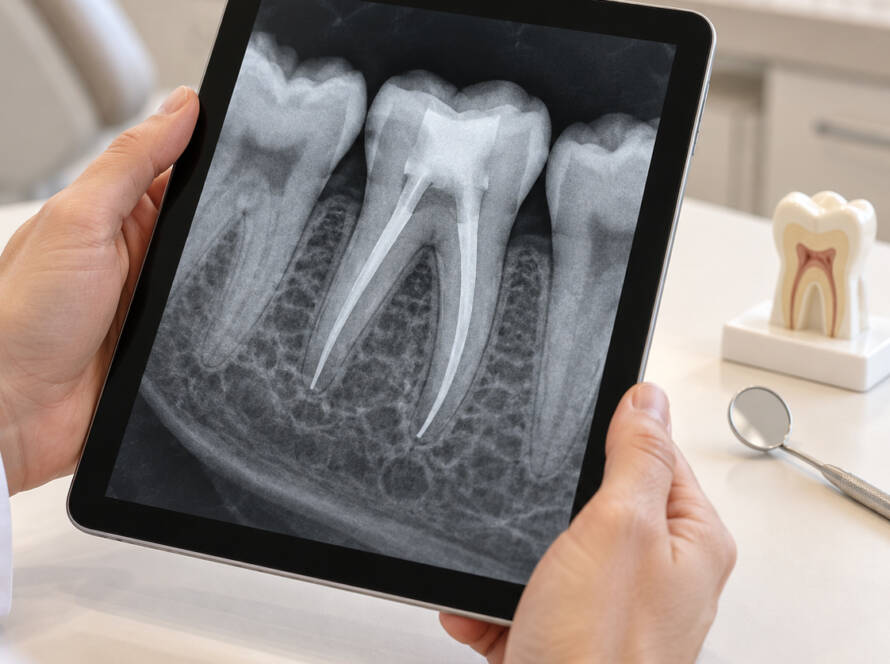
A periapical radiograph shows a tooth or small group of teeth from crown to root tip together with surrounding bone. It can support assessment of local root-area changes, endodontic questions and selected periodontal findings.
Cone-Beam CT
CBCT creates a three-dimensional representation of a selected dental and maxillofacial region. It may be useful when three-dimensional anatomical relationships are important and when the additional information is expected to influence diagnosis or treatment planning.
CBCT is not automatically “better” for every dental question. The imaging method should match the diagnostic need.
How Is Dental Imaging Chosen?
Imaging should follow the clinical question rather than a fixed routine. Before an image is taken, the dentist considers the patient’s symptoms, examination findings, previous radiographs and the treatment being planned. Existing images may already answer the question, while in other cases a different view or an updated image may be justified.
The practical process is therefore: clinical examination, definition of the diagnostic question, selection of the most appropriate imaging method, acquisition of a diagnostically usable image and interpretation together with the clinical findings. The aim is to obtain the information needed for treatment planning without unnecessary repeat exposure.
During image acquisition, positioning and preparation depend on the method. A periapical image, panoramic radiograph and CBCT examination do not involve the same positioning or field of view. Technical adequacy is checked because a poor-quality image that has to be repeated can create unnecessary exposure.
CBCT in Implant and Surgical Planning
Three-dimensional assessment can be important when planning implant placement and evaluating relevant anatomical structures. CBCT may also be considered for selected impacted teeth, complex endodontic questions and some surgical conditions.
The need and field of view should be selected according to the clinical question. A previous image can also be considered when deciding whether new imaging is necessary; repeat exposure should not be performed simply because a patient has travelled to another clinic.
Imaging in Different Areas of Dental Treatment
The role of imaging changes according to the treatment question. In implant planning, imaging can help assess bone and relevant anatomical structures; current ADA/AAOMR recommendations support panoramic imaging for initial assessment and CBCT for presurgical implant planning and placement when three-dimensional site evaluation is required. This does not mean CBCT should be repeated for routine review of an asymptomatic implant.
In endodontics, intraoral two-dimensional radiographs are commonly used for initial assessment, with CBCT reserved for selected cases in which three-dimensional information is expected to change diagnosis or management. In periodontology, radiographs can help assess supporting bone levels alongside periodontal examination. In oral surgery, imaging may clarify the position of impacted teeth and their relationship to nearby structures.
Restorative dentistry can also use radiographs to assess selected caries lesions, existing restorations and areas that cannot be evaluated directly during examination. The imaging method is chosen according to the question; no single modality is automatically required across all of these fields.
Using Existing Dental X-Rays When Travelling for Treatment
Existing dental images can be useful when a patient is seeking treatment in another country, but usefulness depends on whether the image answers the current clinical question. The region shown, image quality, changes that may have occurred since it was taken and the treatment now being considered all matter.
Travelling to a new clinic is not, by itself, a reason to repeat radiographs. A clinician may be able to use previous images, may need a different view, or may decide that updated imaging is justified after examination. For patients, the practical decision is to keep available dental images and relevant records accessible and to ask which records are useful for the planned assessment.
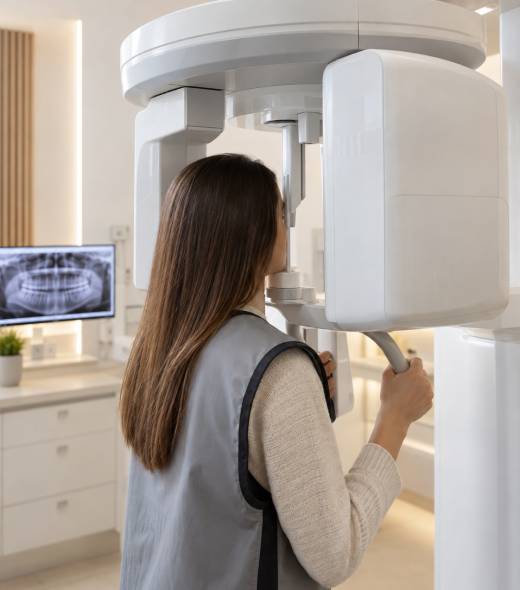
Radiation and Safety
Dental X-rays use ionising radiation. The goal is therefore to obtain clinically necessary information while avoiding unnecessary exposure and optimising the examination.
It is misleading to describe dental imaging as either completely harmless or inherently dangerous. The expected diagnostic benefit and radiation exposure should be considered together.
Pregnancy does not automatically prohibit clinically necessary dental radiography, but pregnancy or possible pregnancy should be communicated to the dental team so that the need for imaging can be assessed appropriately.
Children also require individual image selection rather than a routine age-based schedule. Caries risk, symptoms, tooth development, examination findings and existing images all influence whether imaging is justified. The field and exposure should be limited to what is needed for the clinical question, and CBCT should be reserved for situations in which it provides information that lower-exposure methods cannot adequately supply.
What Happens During an Imaging Appointment?
The time required depends on the imaging method and the area being examined. A local periapical radiograph, a panoramic image and a CBCT examination are different procedures, so a single standard duration is not meaningful.
Before imaging, the relevant symptoms, previous images and treatment question are reviewed. Depending on the method, removable metal objects may need to be taken out and the patient may need to remain in a specific position while the image is acquired. Afterwards, image quality and diagnostic usefulness are assessed before the findings are interpreted with the clinical examination.
What Affects the Cost of Dental Imaging?
Cost depends on the type and scope of imaging that is clinically required. A local intraoral radiograph, panoramic radiography and CBCT do not represent the same technical examination, and CBCT field of view can also vary with the diagnostic question. The appropriate imaging method should be selected first; individual cost can then be determined without assuming that the most extensive examination is necessary.
Imaging at Güler Dent
Güler Dent has in-clinic tomography technology. Three-dimensional imaging is used according to clinical need rather than as an automatic step for every dental treatment.
The clinic’s broader treatment areas include dental implants, endodontics, periodontology, oral and maxillofacial surgery, restorative dentistry and prosthetic dentistry, all of which can involve imaging decisions in selected cases.
Smiles Shaped by Science, Experience, and Trust
Frequently Asked Questions
Do I need CBCT before every dental treatment?
No. Imaging is selected according to the clinical question.
Is CBCT the same as a panoramic X-ray?
No. A panoramic image is two-dimensional; CBCT provides three-dimensional volumetric information.
Is CBCT used for dental implants?
Three-dimensional imaging can be important for implant-site assessment and surgical planning. The exact imaging decision is clinical.
Should dental X-rays be repeated automatically?
No. Existing images, their diagnostic quality and the current clinical need should be considered before new exposure.
Are dental X-rays safe?
They involve ionising radiation, so unnecessary exposure should be avoided. When imaging is clinically justified, the expected diagnostic benefit is weighed against exposure.
Can dental X-rays be taken during pregnancy?
Pregnancy does not automatically rule out necessary dental imaging. The dental team should be informed so the clinical need can be assessed.
Dental Imaging Assessment in Samsun, Turkey
If dental imaging is needed as part of treatment in Turkey, the appropriate examination should be chosen according to the clinical question rather than by routinely repeating X-rays or using CBCT for every patient. If you already have recent dental images or relevant records, keeping them available can help the clinician decide whether they remain useful or whether a different or updated view is justified. To arrange a dental assessment with Güler Dent Ağız ve Diş Sağlığı Polikliniği, contact the clinic at the details below.
- Address: Mimarsinan Mahallesi, Atatürk Boulevard, No: 266, Atakum, Samsun, Turkey
- Phone: +90 552 439 00 01
Imaging selection is made as part of an individual clinical assessment and is based on diagnostic need.
This page provides general information and does not replace a clinical examination or professional radiographic assessment.