

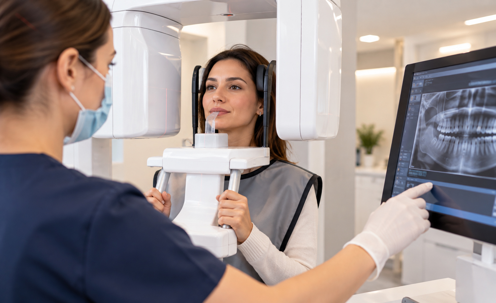
Dental X-rays use ionising radiation, so they are not literally radiation-free. Safety depends on using imaging only when it is clinically justified and choosing the method, field and settings that provide the necessary information without unnecessary exposure.
For most patients, appropriately selected dental radiography provides useful diagnostic information with relatively low radiation exposure. Whether a new image is needed depends on symptoms, examination findings, disease risk, previous radiographs and the treatment being considered.
Pregnancy does not automatically prohibit clinically necessary dental X-rays. Children can also have dental radiographs when indicated, but imaging should be selected according to individual need rather than a fixed schedule.
What “Safe” Means in Dental Radiography
In medical imaging, safety does not mean zero exposure or zero theoretical risk. It means that the expected clinical benefit justifies the exposure and that the examination is optimised to avoid unnecessary dose.
Before taking an image, the dental team considers whether it is expected to answer a specific diagnostic or treatment-planning question, whether a recent image already provides that information, whether a smaller field or lower-dimensional examination is sufficient, and whether the technique and positioning can minimise the need for repeat exposure.
The appropriate image is the one that adds clinically useful information without unnecessary radiation.
X-Rays Are Not Needed at Every Check-Up
There is no universal schedule that requires every patient to have the same set of dental X-rays at every visit.
Someone with active caries, new symptoms or a complex treatment plan may need imaging at a different interval from a patient with stable oral health and useful recent radiographs. Age, stage of dental development, caries and periodontal risk, previous treatment, trauma and planned procedures all influence selection.
Routine habits such as taking a panoramic image every year are not a substitute for clinical justification.
Radiation Dose Varies by Examination
A small intraoral image, a panoramic radiograph and a CBCT scan are not equivalent examinations. Radiation dose varies according to imaging type, equipment, field of view, technique and acquisition protocol.
Because device and protocol differences can be substantial, a single dose number can be misleading when it is presented as though it applies to every clinic and patient.
The more useful question is whether the selected examination is appropriate for the diagnostic information needed.
Dental X-Rays During Pregnancy
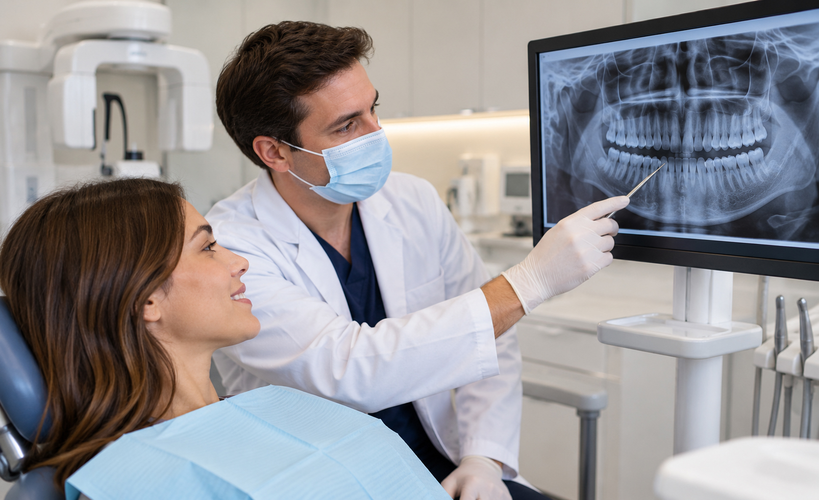
Current professional guidance does not treat pregnancy as an automatic contraindication to clinically necessary dental radiography. If an image is needed to diagnose or treat a dental problem, pregnancy alone is not a reason to leave the condition unassessed.
Patients should tell the dental team if they are pregnant or think they may be pregnant so that this information can be included in the clinical decision.
Unnecessary radiographs should be avoided during pregnancy just as they should be avoided in other patients. Necessary dental imaging, however, is not automatically forbidden. Delaying diagnosis of significant pain or infection simply because an image is required can also create avoidable health problems.
Dental X-Rays for Children
Children can have dental radiographs when clinically indicated. Because they are still growing and have a longer lifetime in which radiation effects could theoretically accumulate, avoiding unnecessary exposure is particularly important.
Imaging may be useful for caries that cannot be assessed fully by examination, tooth development and eruption, dental trauma, orthodontic assessment, suspected infection or unerupted and missing teeth.
The image should be selected according to the child’s age, size, symptoms and diagnostic need rather than a routine timetable.
Panoramic X-Ray and CBCT
A panoramic radiograph is a two-dimensional overview, while CBCT provides three-dimensional information.
CBCT can be justified when spatial anatomy, bone dimensions, impacted-tooth relationships or complex endodontic questions require 3D assessment. It is not automatically safer or better simply because it is newer or more detailed.
If a panoramic or intraoral image answers the clinical question, additional CBCT may not be necessary.
Repeating Images at a New Clinic
Changing clinics or travelling to another country does not, by itself, justify repeating dental imaging.
Existing images can remain useful if they include the relevant area, are of diagnostic quality and are appropriate for the current treatment question. A new image may still be needed if the existing examination is outdated for the problem, does not cover the correct region or lacks sufficient diagnostic quality.
Sharing previous radiographs where possible can help avoid unnecessary duplication.
Lead Aprons and Thyroid Collars
Current ADA radiation-safety recommendations no longer advise routine use of abdominal lead aprons or thyroid collars for modern dental imaging, including for pregnant patients and children, when the examination is performed correctly.
Modern collimation, suitable exposure settings, accurate positioning and avoiding unnecessary examinations provide more meaningful protection. Shielding can sometimes interfere with the primary beam and lead to repeat imaging.
Local regulations and institutional policies can differ, so patients should follow the imaging facility’s current protocol.
How Dental Teams Reduce Unnecessary Exposure
Radiation safety depends on the entire imaging process. Important measures include taking radiographs only when clinically indicated, using the smallest appropriate field of view, selecting suitable imaging parameters, positioning the patient correctly, reviewing existing images before repeating an examination and using 2D imaging when it is sufficient.
Good radiology begins with the clinical question rather than with the equipment available.
Dental Imaging at Güler Dent
Dental Radiology is one of the main clinical service areas at Güler Dent in Samsun, Turkey. Güler Dent has in-clinic tomography technology for cases in which three-dimensional imaging is clinically indicated.
The presence of CBCT technology does not mean every patient receives a scan. Imaging is selected according to symptoms, examination findings and the diagnostic information required.

