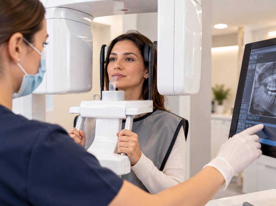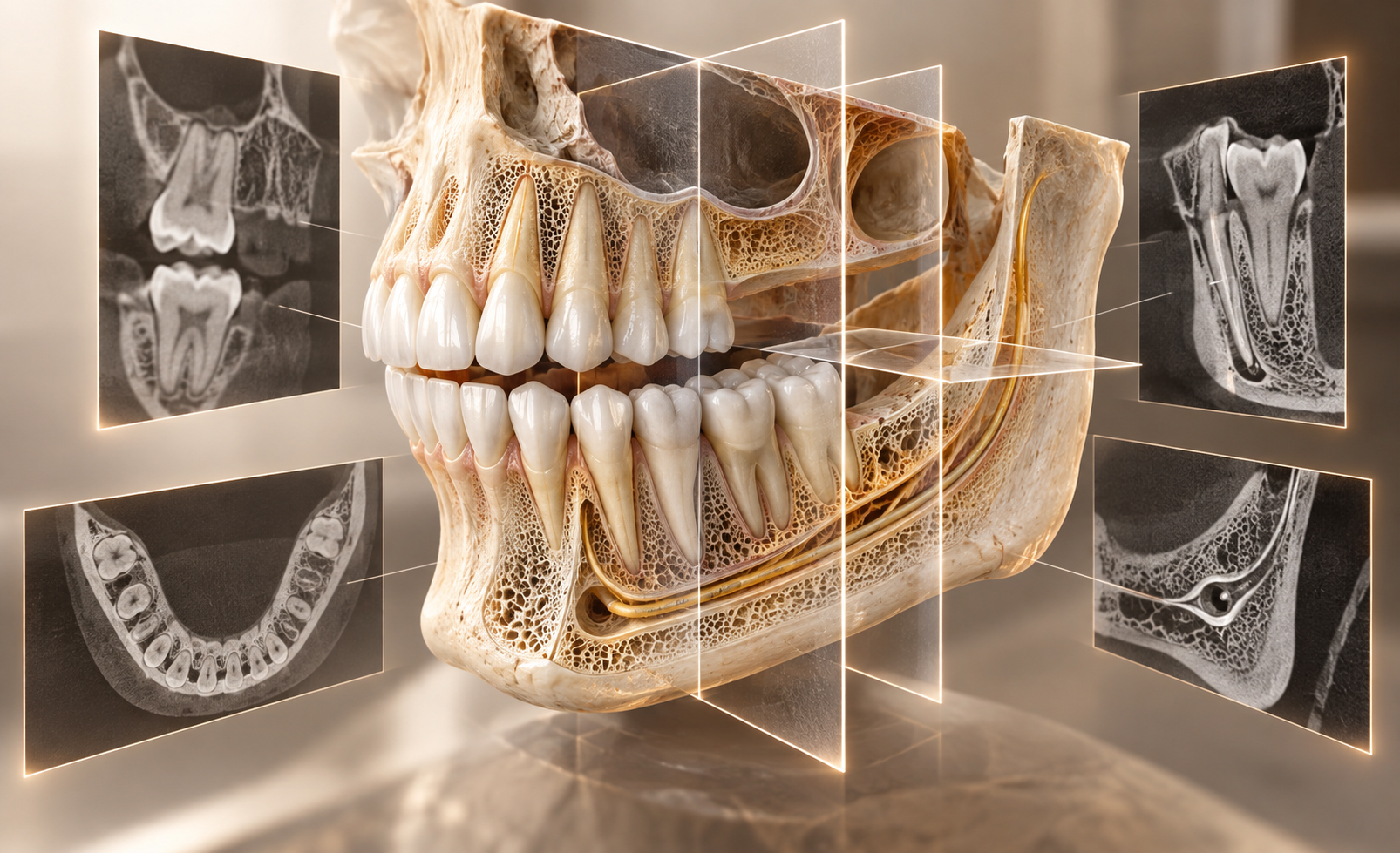
A panoramic dental X-ray and a dental CBCT scan answer different types of questions. A panoramic radiograph provides a broad two-dimensional view of the teeth and jaws, while CBCT creates a three-dimensional volumetric dataset that can be examined in multiple planes and cross-sections.
CBCT is particularly useful when a dentist needs to understand bone dimensions, the spatial relationship between a tooth and nearby anatomy, or complex root or surgical anatomy that cannot be assessed adequately in 2D. That does not make it the default or “better” image for every patient. If a clinical question can be answered with an appropriate 2D image, additional 3D imaging may not be justified.
The Main Difference Is Dimensional Information
A panoramic radiograph compresses curved three-dimensional anatomy into a single 2D image. It provides wide coverage but does not show true depth or cross-sectional bone dimensions.
CBCT acquires data from multiple projections and reconstructs a three-dimensional volume. The clinician can review the area in axial, coronal, sagittal and other reconstructed views, which can clarify anatomy hidden by overlap on a conventional 2D image.
The extra information is valuable only when it answers a real clinical question.
When Panoramic Imaging Is Useful
Panoramic radiography is well suited to situations that require a broad overview. It can help assess tooth development and eruption, impacted or partially impacted teeth, broad jaw anatomy, larger bony changes and general relationships between teeth and surrounding structures.
It is also useful as a first-line overview before deciding whether more focused imaging is needed. Its wide field, however, does not make it ideal for every small lesion or detailed root question.
What CBCT Adds
The main advantage of CBCT is depth and cross-sectional information. Depending on the field of view and image quality, it may help assess bone width and height in a selected region, the three-dimensional relationship to the mandibular canal or maxillary sinus, impacted teeth in relation to nearby structures and complex root anatomy.
In selected endodontic situations it can also help investigate resorption, suspected missed canals, some fractures or previously treated teeth with persistent disease.
The value comes from answering a specific question, not from collecting more data for its own sake.
Implant Planning
Three-dimensional information can be important in implant planning because implant position depends on available bone and its relationship to anatomical structures. CBCT can provide cross-sectional assessment of the site and support prosthetically driven surgical planning.
A CBCT scan does not determine implant candidacy by itself. Periodontal health, soft tissues, restorative space, general health and the final prosthetic plan still need clinical assessment.
Imaging should therefore be selected as part of treatment planning rather than applied automatically to every missing tooth.
Wisdom Teeth and Surgical Planning
A panoramic radiograph provides enough information for the initial assessment of many wisdom teeth.
CBCT may be considered when the 2D image suggests a close or uncertain anatomical relationship and three-dimensional information could change the surgical plan. A common example is an impacted lower third molar that appears close to the inferior alveolar nerve canal.
The deciding factor is whether the additional 3D information is likely to influence management.
Selective Use in Endodontics
Periapical radiographs remain important in routine endodontic assessment. CBCT is reserved for situations in which two-dimensional imaging does not explain the clinical problem adequately, such as complex anatomy, suspected missed canals, selected fractures, resorption or persistent disease in a previously treated tooth.
It should not be used as a screening examination for every root canal.
CBCT Does Not Replace Conventional Dental X-Rays
Three-dimensional imaging does not make bitewings, periapical radiographs or panoramic X-rays obsolete.
Bitewings are particularly useful for selected caries and bone-level questions. Periapical radiographs provide detailed local views of teeth and roots, while panoramic imaging gives a broad overview.
Choosing the right modality means matching the image to the task. More dimensions do not automatically produce more diagnostic value.
Field of View and Why It Matters
Field of view, or FOV, describes the volume included in a CBCT scan. A smaller field can focus on a limited region, while a larger field includes more anatomy.
When CBCT is justified, the selected field should be appropriate for the diagnostic question rather than automatically exposing a broader region than necessary. Field of view, device settings, image resolution and acquisition protocol all affect radiation dose and image quality.
Radiation and Justification
Both panoramic radiography and CBCT use ionising radiation. CBCT dose varies widely according to the machine, field of view and acquisition protocol, so a single universal dose number can be misleading.
Current dental imaging practice is based on justification and optimisation: use radiographs when they are expected to provide clinically useful information, select the appropriate modality and settings, and avoid unnecessary repeat exposures.
CBCT should not be chosen simply because the equipment is available.
How the Imaging Decision Is Made
The process starts with the clinical question. The dentist considers what condition is being investigated, whether a broad overview or local detail is needed, whether 3D spatial information could change diagnosis or treatment, whether suitable existing images are available and whether a lower-dimensional image already answers the question.
If CBCT is needed, the field of view should be chosen to match the area and purpose of the examination.
CBCT at Güler Dent
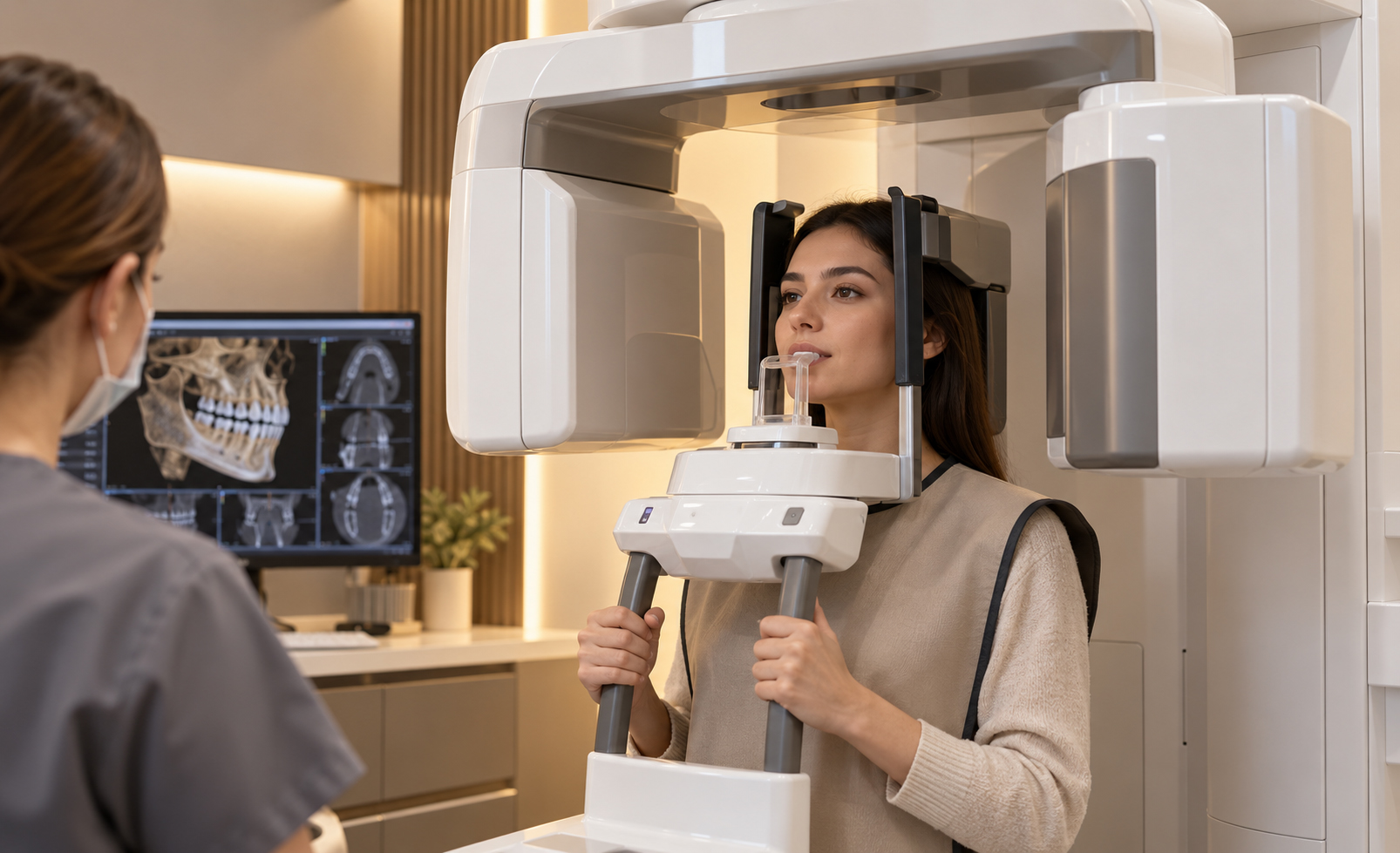
Dental Radiology is one of the main clinical service areas at Güler Dent in Samsun, Turkey. Güler Dent has in-clinic tomography technology.
Three-dimensional imaging is used according to clinical need rather than as a routine step for every patient. CBCT may support selected implant, surgical or endodontic assessments when the additional spatial information is expected to affect planning.